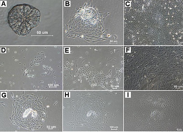
Figure 1. Proliferation of primary-cultured fetal mouse intestinal epithelial cells obtained using different enzymatic methods. Crypts were isolated from fetal mouse intestines using collagenase I/hyaluronidase digestion (A). Proliferative epithelial cells gradually migrated out around the crypts within 24 h (B), formed large colonies after 5 days (C), and continued to spread extensively before confluence was reached. While trypsin digestion yielded mostly single cells, only part of epithelial cells were adhesive and grew slowly (D). Epithelial cells were mixed with fibroblasts (white arrows), which grew either in groups or scattered (E, F). In addition, thermolysin also appeared to give a few crypts, although mostly in single cells. Proliferative epithelial cells migrated outward after 24 h, and spread extensively and formed colonies after culturing for 2 to 6 days (G, H). However, the colonies later stopped expanding, and part of those cells began to degenerate after 10 days (I).

