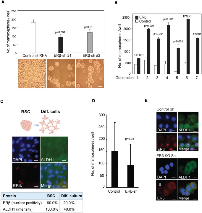
Figure 2.
Estrogen receptor (ER)β and the cancer stem cell phenotype. A) Average number of mammospheres in control vs shRNA-ERβ knockdown clones generated from 2000 MCF7 cells after seven days of incubation in nonadherent conditions, with representative images presented below each bar (Student’s t test, mean ± SD, 3 replicates). Scale bar = 100 µm. B) Number of mammospheres formed by MCF7 cells, with transduced ERβ expression (black bar) and without (white bar) over seven passages. An equal number of cells was seeded for each passage (Student’s t test, mean ± SD, 3 replicates). C) Forced differentiation of breast cancer cells with tumor-initiating capabilities by incubation in selective medium supplemented with 5% fetal bovine serum induced a switch from nonadherent to a spindle-like, adherent cell phenotype and stained for ERβ and ALDH1 (n = 8 patients). Scale bar = 10 µm. D) Numbers of patient-derived mammospheres after lentiviral shRNA-mediated knockdown of ERβ compared with treatment with the nontargeted scrambled shRNA construct as control. Following lentiviral transduction, cells were incubated in selective medium for seven days (Student’s t test, mean ± SD, n = 4 patients, 4 replicates). E) Immunofluorescence imaging of patient-derived cells after lentiviral shRNA-mediated knockdown of ERβ, with antibodies for ERβ (red) and ALDH1 (green) counterstained with DAPI (blue). Scale bar = 10 µm. All statistical tests were two-sided. ALDH1 = Aldehyde dehydrogenase 1; BSC = breast cancer cells with tumor-initiating capabilities; DAPI = 4’,6-diamidino-2-phenylindole; ER = estrogen receptor; sh = small hairpin RNA.

