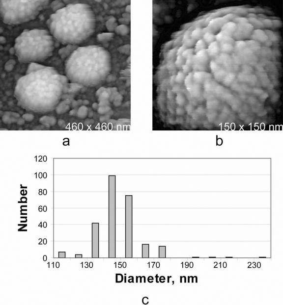
FIG. 1.
(a) AFM image of MuLV virions isolated from culture media and spread on poly-l-lysine-coated glass substrates. (b) A single virion at higher magnification. The appearance of the particles is essentially the same as that of particles associated with host cell surfaces (6). The tufted protein arrangement seen on the virion surfaces is characteristic of both MuLV and HIV. (c) Histogram of particle sizes (corrected for shrinkage due to dehydration) for wild-type MuLV isolated from the culture media of virus-infected NIH 3T3 cells (strain 43-D) and spread on glass substrates for analysis by AFM. The mode of the distribution is 145 nm.

