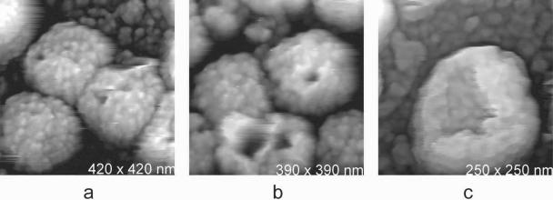
FIG. 3.
(a and b) MuLV particles on glass substrates which exhibit deep pits on their surfaces. These are likely due to loss of surface proteins or sectors of proteins incurred in the preparation process. Similar defects have, however, also been seen on particles still associated with host cell surfaces (6). (c) The shell remaining after loss of the nucleocapsid from an MuLV virion was produced by centrifugation during sample preparation. The shell retains its structural integrity in spite of the loss of a large sector of its surface.

