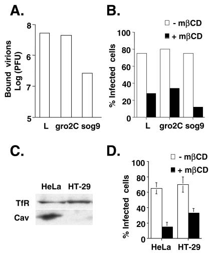
FIG. 7.
mβCD blocks WT VV infection in GAG-deficient and caveolin-deficient cells. (A) L, gro2c, and sog9 cells were infected with WT VV at an MOI of 10 at 4°C for 30 min, washed, and then harvested. The amounts of bound IMV were determined by plaque assays with BSC40 cells. (B) Cells were treated with medium alone (−mβCD) or medium containing 10 mM mβCD (+mβCD) and subsequently infected with vMJ360 at an MOI of 5 PFU per cell (for L and gro2 cells) or 20 PFU per cell (for sog9 cells). The cells were fixed at 2 h p.i. and stained with X-Gal, and the numbers of blue cells were counted. The percentage of infected cells = 100 × [(number of blue cells)/(number of blue cells + number of white cells)]. (C) Immunoblots of HeLa and HT-29 cell lysates with anti-transferrin or anti-caveolin Ab. (D) HeLa and HT-29 cells were treated with medium alone (−mβCD) or medium containing 10 mM mβCD (+mβCD) and subsequently infected with vMJ360 at an MOI of 10 PFU per cell. Viral early gene expression was determined by a β-Gal staining assay as described for panel B.

