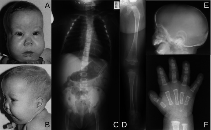
Figure 1.

Clinical and radiological features of the patient at 7 months of age. (A, B) Facies with flat nasal root, epicanthic folds, long philtrum, and micrognathia; macrocephaly and low‐set ears. (C–F) Skeletal survey. (C) Hypoplastic scapulae, cervical kyphosis, thoracic scoliosis with undermineralized thoracic pedicles, 11 pairs of ribs, short ischia and unossified inferior pubic rami. (D) Lower limb with straight femur, tibia, and fibula, with delayed ossification of the femoral epiphysis. (E) Lateral skull film showing enlargement of the cranial vault to the size of the facial bones. (F) Hand with short metacarpal I and moderately short middle and distal phalanges.
