
Figure 2.
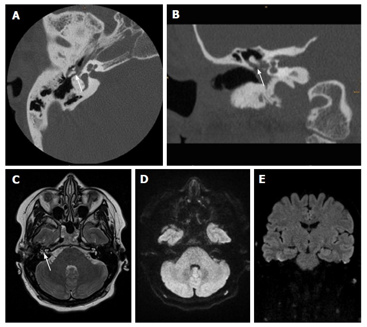
Thirty-one-year-old female after surgery for cholesteatoma. A, B: CT show a soft-tissue mass in the tympanic space adjacent to malleolus and scutum with suspected bony erosion (white arrow); C-E: Axial T2 weighted image shows fluid-like signal (white arrow) that has no restriction in EPI DWI RESOLVE (axial in D and coronal in E). There was no sign of recurrent cholesteatoma on follow-up surgery. CT: Computed tomography; DWI: Diffusion weighted imaging; EPI: Echo-planar imaging; RESOLVE: Readout-segmented echo-planar.