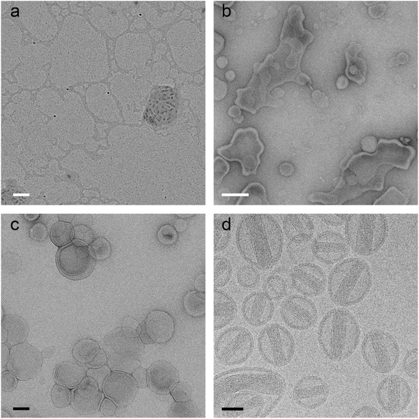
Figure 5.

Doxorubicine liposomes (Dox‐NP®) imaged by the most frequently used techniques in soft matter electron microscopy: dried sample without staining (a), UAc stained sample after two minutes of drying (b), negative stained sample (UAc) (c) and cryo‐TEM (d). White scale bars represent 200 nm and black scale bars 50 nm.
