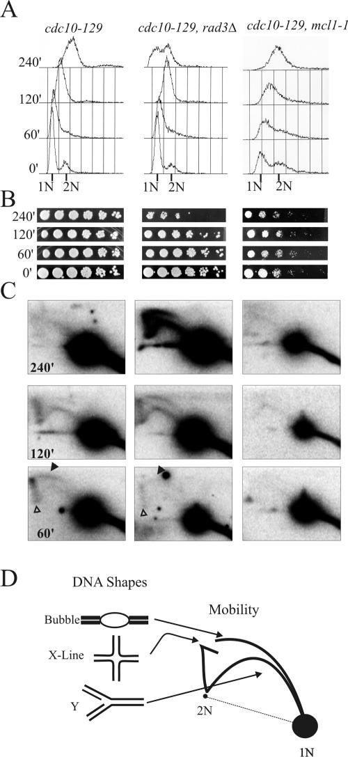
FIG. 4.
DNA replication forks at ars2-1 in mcl1-1 mutants examined during replication stress are stable and persistent. The three cdc10-129 strains shown in Fig. 3 were arrested at 36°C and released into 5 mM HU to induce replication stress. Genotypes of strains are given at the top of each column, and each row is labeled from the corresponding time points. (A) Flow cytometric analysis of DNA content in cells released into 5 mM HU. Cytology taken through the experiment showed that most cells entered mitosis by 240 min, except for mcl1-1 cells, which appeared cell cycle arrested at this time point (data not shown). (B) Relative viability was assessed in serial plating of cultures at each time point. (C) Southern blot analysis of 10 μg of total DNA by two-dimensional gels probed for ars2-1 sequences (an early firing origin). Quantification of total signal with Molecular Dynamic ImageQuant 5.1 shows that the hybridization signal increases twofoldin the cdc10-129 (3 × 10⁵ to 6 × 10⁵ counts) and rad3Δ (3 × 10⁵ to 5.5 × 10⁵ counts) strains but not the mcl1-1 strain (2 × 10⁵ to 2.4 × 10⁵ counts). Filled arrowheads mark the bubble arc, and open arrowheads mark the X line. (D) Diagrammatic representation of two-dimensional DNA gel patterns showing how autonomously replicating sequence DNA structure relates to mobility in two-dimensional gel electrophoresis, which is adapted from http://fangman-brewer.genetics.washington.edu/2Dgel.html.

