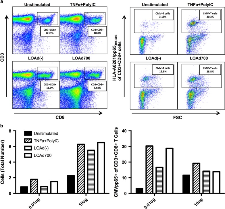
Figure 6.
LOAd700-stimulated DCs expand antigen-specific T cells and NK cells. Peripheral blood mononuclear cells expanded using DCs from CMV-positive blood donors (n=3). (a) Expansion of CD3+, CD8+ and double-positive T lymphocytes of all cultured cells are shown in the left panel, and the percentage of CMV-specific T cells of all gated CD3+CD8+ T cells against forward scatter are shown in the right panel. (b) The left panel demonstrates the total number of cells in the co-cultures using 0.01 or 10ug pp65 CMV peptides. The right panel shows percentage of CMV-specific CD3+CD8+ T cells in the co-cultures using 0.01 or 10 μg pp65 CMV peptides as calculated by multiplying the % cells with the manual count of cells in the cultures. Representative figures are shown.

