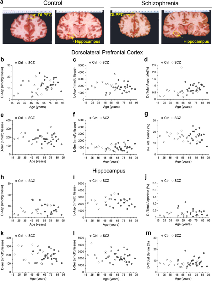
Fig. 1.

Age-related changes in d-aspartate and d-serine levels between patients with schizophrenia and control subjects in the post-mortem dorsolateral prefrontal cortex and hippocampus. a Representative images of the dorsolateral prefrontal cortex (DLPFC) and hippocampus dissected from post-mortem brains of healthy (control, Ctrl) and schizophrenia-affected individuals (SCZ). The dot plots represent the variations across lifespan in the content of free b, h d-aspartate and e, k d-serine, c, i l-aspartate and f, l l-serine, and d, g, j, m d-/total amino acids in the b–g dorsolateral prefrontal cortex (DLPFC) and h–m hippocampus of non-psychiatric individuals (DLPCF, n = 20; hippocampus, n = 15) and patients with schizophrenia (DLPCF, n = 19; hippocampus, n = 15). In each sample, all the amino acids were detected in a single run by HPLC and expressed as nmol/g of tissue, while the ratios are expressed as percentage (%)
