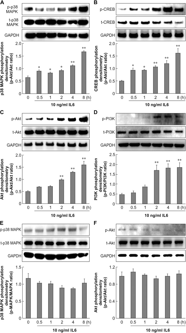
Fig 6. Effect of IL-6 on p38 MAPK and Akt signaling in primary skin fibroblasts.
Primary skin fibroblasts from control mice (A, B, C, D) and diabetic mice (E, F) were serum-starved for 16 h. Cells were treated with 10 ng/mL IL-6 and incubated for 0.5, 1, 2, 4, and 8 h. A: Cell lysates were analyzed by western blotting using anti-phospho-p38 MAPK and anti-p38 MAPK antibodies. The data represent the mean ± SE of 3 different experiments (*: p < 0.05, **: p < 0.01 vs. 0 h). B: Cell lysates were analyzed by western blotting using anti-phospho-CREB and anti-CREB antibodies. The data represent the mean ± SE of 3 different experiments (*: p < 0.05, **: p < 0.01 vs. 0 h). C, D: Cell lysates were analyzed by western blotting using anti-phospho-Akt, anti-phospho-PI3K, anti-Akt, and anti-PI3K antibodies. The data represent the mean ± SE of 3 different experiments (**: p < 0.01 vs. 0 h). E, F: DM cell lysates were analyzed by western blotting using anti-phospho-p38 MAPK, anti-phospho-Akt, anti-p38 MAPK, and anti-Akt antibodies. The data represent the mean ± SE of 3 different experiments.

