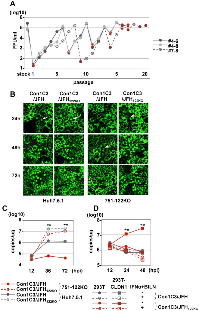
Fig 7. Propagation of Con1C3/JFH122KO in 751-122KO cells.
(A) Infectious titer in the culture medium on serial passage of 751-122KO#1 or Huh7.5.1 cells. Red circles indicate the passage in 751-122KO cells, and the other circles indicate the passage in Huh7.5.1 cells. Three independent passages (#4–6, #4–8, #7–8) are shown. (B) Nuclear translocation of IPS-GFP (arrows) in Huh7.5.1 and 751-122KO cells upon infection with Con1C3/JFH and Con1C3/JFH122KO. (C) Con1C3/JFH and Con1C3/JFH122KO were inoculated into 751-122KO#1 and Huh7.5.1 cells, and the levels of intracellular HCV-RNA replication were determined. Error bars indicate the standard deviation of the mean and asterisks indicate significant differences (**P < 0.01) versus the results for the control. (D) 293T-CLDN cells infected with either Con1C3/JFH or Con1C3/JFH122KO were treated with IFNα and BILN and then the intracellular HCV-RNA level was determined at 12, 24 and 48 hpi. Error bars indicate the standard deviation of the mean and asterisks indicate significant differences (**P < 0.01) versus the results for the control.

