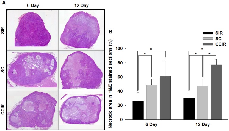
Fig 5. Histologic analysis of the tumor response.
(A) The mean percentage tumor necrotic fraction was determined from hematoxylin and eosin- (H&E) stained sections after ionizing radiation (IR), celastrol, and combined IR celastrol therapy at day 6 and 12 after treatment initiation (original magnification, ×100). (B) Graph of the percentage necrotic area in H&E-stained sections. Tumors treated with the combined IR and celastrol therapy showed a significantly (p = 0.05) larger necrotic area at day 6 and 12 than that observed in the IR and celastrol mono-treatment groups. *p = 0.05 (statistically significant). SIR, single ionizing radiation; SC, single celastrol; CCIR, celastrol-combined ionizing radiation.

