
Figure 2.
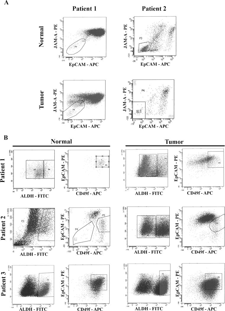
Flow cytometry sorting of cells for single cell analyses. A) Jam-A/EpCAM staining to separate breast epithelial cells from feeder layer fibroblasts. Fibroblasts do not stain for Jam-A/EpCAM. Jam-A/EpCAM positive cells were sorted and used for unselected cell analyses. B) Sorting of ALDH+/CD49f+/EpCAM+ cells to enrich for phenotypically defined cell population. CD49f+/EpCAM+ cells in the boxed regions on right were selected for analyses.