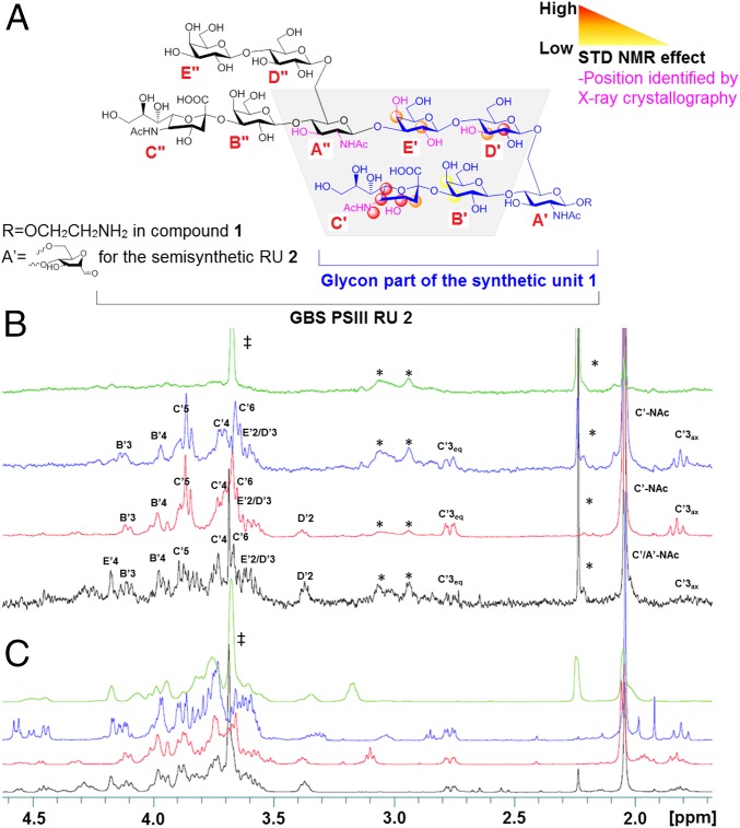
Fig. 3.
GBS PSIII epitope mapping. (A) Protons interacting with the mAb identified by STD-NMR and X-ray crystallography. (B) STDD and (C) related 1H NMR spectra of the desialylated tetrasaccharide [β-Gal-(1→4)–β-GlcNAc-(1→3)–β-Gal-(1→4)–α/β-Glc in green], the linear 3 (blue) and branched 1 (red) RUs, and the DP2 fragment (black). Proton positions receiving saturation after irradiation of the protein are indicated. ‡ indicates the CH2 signal of the Tris buffer; * refers to signals related to the protein.

