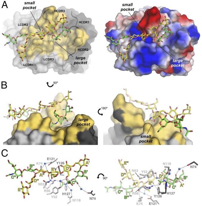
Fig. 4.
Crystal structure of DP2 bound to Fab NVS-1-19-5. (A, Left) The Fab is depicted with surfaces and the DP2 with sticks. Fab residues involved in direct binding with DP2 (the paratope) are colored in yellow, and the binding pockets are labeled. (A, Right) Surface electrostatic potential distribution of the Fab, oriented as on Left. (B) Two views (Left and Right) of the large and small pockets where the DP2 binds. (C) Details of the interactions between DP2 and Fab. Carbon atoms of the DP2 backbone are colored in yellow, and those belonging to the branches are in green, whereas carbon atoms of the Fab are colored in light and dark gray for the L and H chain, respectively. Nitrogen and oxygen atoms are colored in blue and red, respectively, and water molecules are shown as blue spheres.

