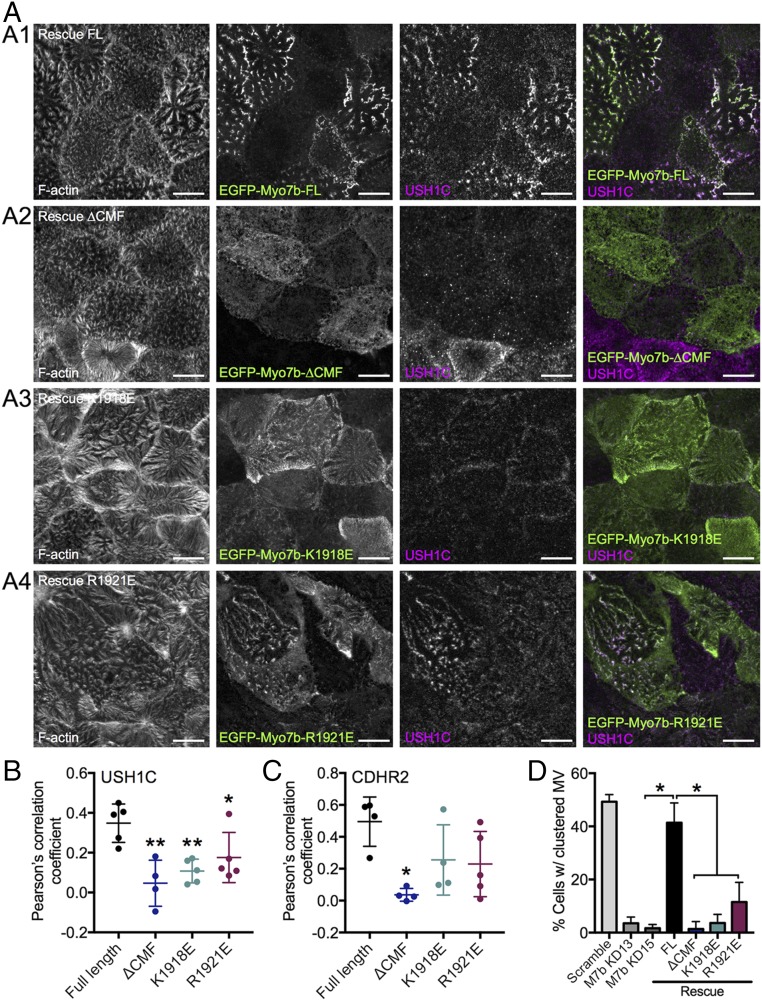
Fig. 3.
Myo7b variants deficient in USH1C binding exhibit decreased colocalization with USH1C and impaired microvillar clustering. (A) Confocal images of 14 DPC Myo7b KD CACO-2BBE cells expressing the EGFP-tagged Myo7b rescue constructs indicated (green) and stained for USH1C (magenta) and F-actin (gray). (Scale bars, 10 μm.) (B) Colocalization analysis of Myo7b with USH1C using Pearson’s correlation coefficients. n = 5 fields, 111 cells for FL; 4 fields, 30 cells for ΔCMF; 5 fields, 66 cells for K1918E; 5 fields, 84 cells for R1921E. *P < 0.05, **P < 0.01, t test. (C) Colocalization analysis of Myo7b constructs with CDHR2 using Pearson’s correlation coefficients. n = 4 fields, 59 cells for FL; 4 fields, 37 cells for ΔCMF; 4 fields, 46 cells for K1918E; 5 fields, 61 cells for R1921E. *P < 0.01, t test. The representative images are shown in Fig. S3. (D) Quantification of cells with clustering microvilli expressed as a total percentage of cells. For KD rescue cell lines, only EGFP+ (i.e., rescue construct-expressing) cells were scored. Bars indicate mean ± SD. n = 606 cells for scramble, 573 cells of Myo7b KD13 experiment, 507 for Myo7b KD15, 271 for FL, 89 for ΔCMF, 261 for K1918E, and 264 for R1921E. *P < 0.002, t test. KD13 and KD15 represent two distinct short hairpin RNA KD lines as described previously (37).

