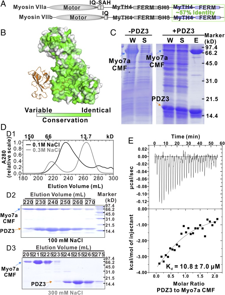
Fig. 4.
Interaction between Myo7a CMF and USH1C PDZ3. (A) Domain organizations of Myo7a and Myo7b. Sequence identity of their CMF is indicated. (B) The amino acid sequence conservation map of Myo7a/b CMF showing that the residues forming the USH1C PDZ3 binding surface are nearly identical. The conservation map is calculated based on amino acid sequences of mammalian Myo7a and Myo7b. (C) SDS/PAGE analysis showing that coexpression of USH1C PDZ3 can increase the solubility of Myo7a CMF. “W” means whole-cell extract; “S” denotes supernatant after centrifugation of the cell lysate; “E” stands for the fraction after elution from the Ni2+-NTA column. (D) The elution profiles of Myo7a CMF/USH1C PDZ3 complex in the buffer containing 100 mM NaCl (black) or 300 mM NaCl (gray). The elution positions of molecular size markers are indicated at the top (D1). The corresponding SDS/PAGE analysis of the eluted peaks (D2: in buffer containing 100 mM NaCl and D3: in buffer containing 300 mM NaCl). (E) ITC result showing that Myo7a CMF binds to USH1C PDZ3 with a Kd ∼11 μM.

