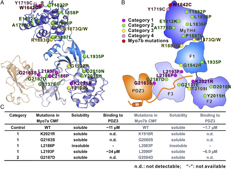
Fig. 5.
Missense mutations in Myo7a CMF found in USH1B patients. (A) Ribbon representation of the Myo7a CMF structural model. The 20 missense mutation sites in USH1B patients are highlighted with spheres and colored in magenta, green, yellow, and pink, corresponding to the four categories. A de novo mutation found in Myo7b (W1642C) is shown in red. (B) Schematic cartoon diagram showing the distributions of the USH1B mutations mapped on to the structural model of Myo7a CMF. (C) Summary of impacts of some USH1B mutations on the Myo7a CMF/USH1C PDZ3 interaction. The matching mutations were also made in Myo7b CMF corresponding to each of the USH1B mutations, and their impacts on USH1C PDZ3 binding are measured.

