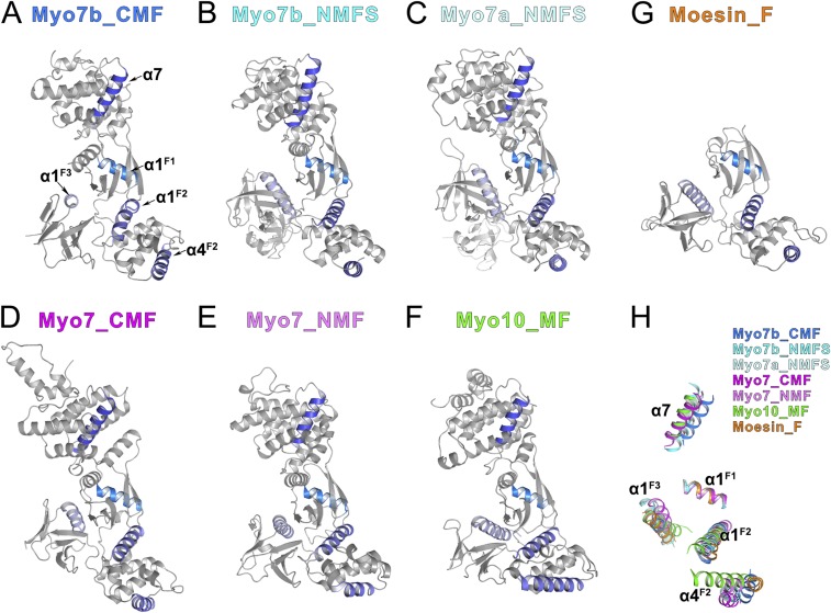
Fig. S1.
Structural comparison of all of the MyTH4-FERM structures showing the relative rotations of the three lobes. (A–G) Ribbon diagrams showing all of the MyTH4-FERM structures solved up to date as well as the founding member Moesin FERM domain. (A) Myo7b CMF, this study; (B) Myo7b NMF, PDB ID code: 5F3Y; (C) Myo7a NMF, PDB ID code: 3PVL; (D) Dictyostelium Myo7 CMF, PDB ID code: 5EJR; (E) Dictyostelium Myo7 NMF, PDB ID code: 5EJY; (F) Myo10 MF, PDB ID code: 3PZD; (G) Moesin FERM domain, PDB ID code: 1EF1. All of the structures are shown in the same orientation by aligning the F1 lobes together. The α7 in the MyTH4 domain, α1 in the F1 lobe, α1/α4 in the F2 lobe, and α1 in the F3 lobe are highlighted in color to show the relative orientations of each domains. (H) Superposition of the five helices highlighted in A–F showing the orientation differences of the MyTH4-FERM tandems using the F1 lobe α1 (α1F1) helix as the reference.

