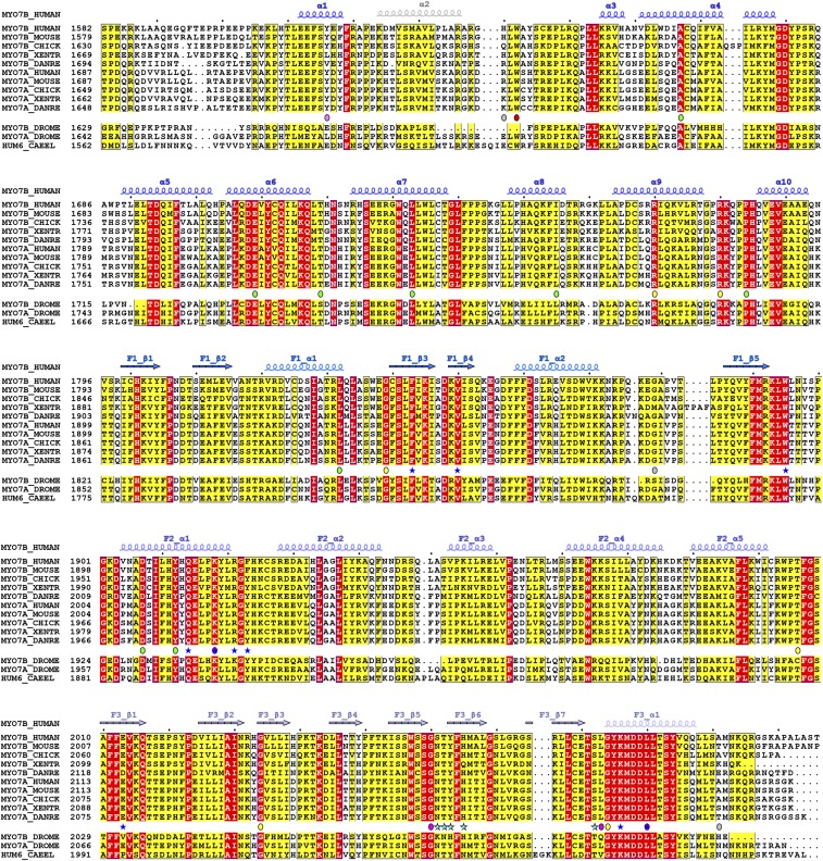
Fig. S4.
Sequence alignment of CMF of class VII myosins from difference species. The secondary structure elements are labeled according to the Myo7b CMF structure. Residues that are identical and highly similar are indicated in red and yellow boxes, respectively. The residues involved in USH1C C-terminal tail binding and the PDZ3 domain binding are highlighted with blue and cyan stars. Missense mutations are highlighted with circles and color coded as that in Fig. 5.

