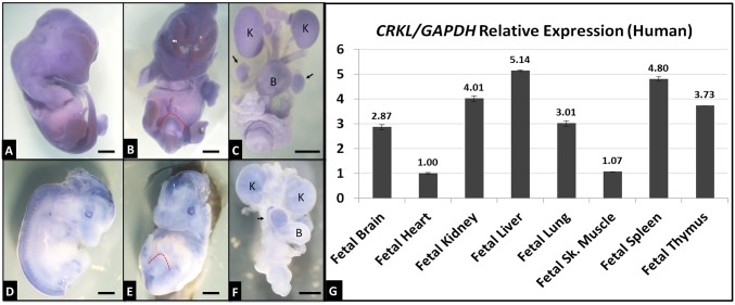
Fig. 2.
Murine Crkl and human CRKL expression patterns. Crkl antisense (A–C) and sense control (D–F) probes were used to stain E12.5 (A, B, D, and E) whole embryos and E16.5 isolated GU tracts (C and F). Blue areas indicate probe hybridization. Crkl is expressed at moderate levels (higher than sense probe background) throughout the developing embryo, including genital tubercle (red dotted outline), kidneys (K), bladder (B), and testes (black arrows). (Scale bars, 2 mm.) CRKL qPCR was performed on cDNAs (G) from spontaneously aborted human fetuses. Expression levels of tissues shown relative to heart expression. Error bars represent SEM.

