Fig. 7.
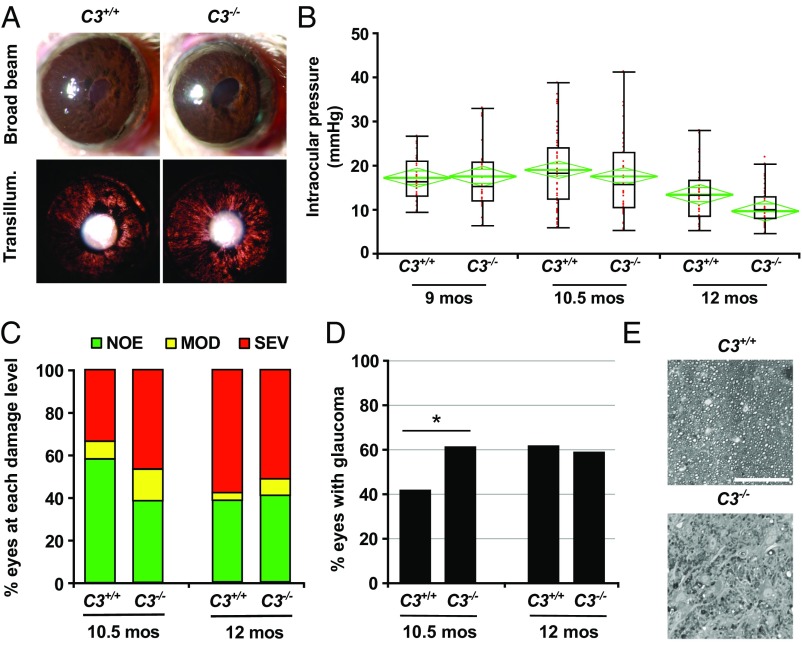
C3 is protective in early D2 glaucoma. (A) D2.C3−/− mice developed iris disease without difference to standard D2 mice. Depigmentation of the iris was observed both by broad-beam illumination (Upper) and transillumination (Lower). (B) Age-dependent elevation of IOP was observed in both C3-sufficient D2 (C3+/+) and C3-deficient D2 (C3−/−) mice, present in a subset of eyes at age 9 mo. There were no significant differences between genotypes at each age assessed (8.5–9.0 mo, P = 0.88; 10–10.5 mo, P = 0.92; 12–12.5 mo, P = 0.94). Boxplots were generated using JMP version 7.0. The boxes define the 75th and 25th percentiles, and the black line in the middle of each box represents the median value. The whiskers depict the full range of the data points. The green diamonds indicate the mean value and the 95% CI. (C and D) Optic nerve damage was significantly increased in C3−/− mice compared with C3+/+ mice at age 10.5 mo. Bars represent the distribution of optic nerve damage by genotype and age. At age 10.5 mo, C3−/− mice exhibited significantly increased nerve damage (P = 0.01). (E) Examples of the most common damage level for C3+/+ (NOE) and C3−/− (SEV) at age 10.5 mo. (Scale bar: 50 μm.)

