Fig. 3.
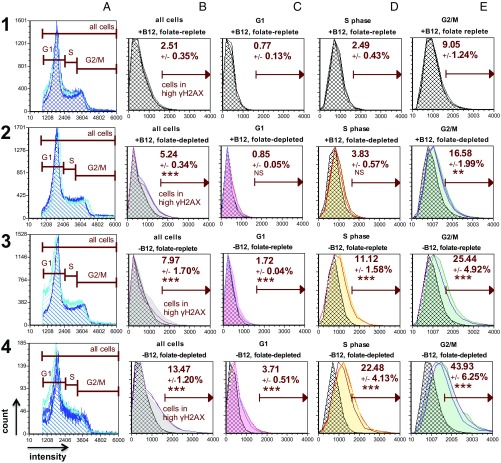
Vitamin B12 depletion induces changes in γH2AX, a marker of DNA damage in HeLa cells. Cells were stained for DNA content (Vybrant Violet; 1–4, A) and γH2AX (FITC; 1–4, B–E); individual plots depict the cell count (y axis) vs. fluorescence intensity (x axis). The high γH2AX parameter is a threshold defined by the mean top 2.5% of cells in G1, S, and G2/M (all cells) stained for γH2AX in the vitamin B12- and folate-replete condition (1, B), and this gate was uniformly applied to all conditions and cell cycle phases. Each plot shows the mean percent high γH2AX ± SD for triplicate measurements for each experimental condition and cell cycle phase. Individual triplicate measurements for each condition are plotted relative to the corresponding mean γH2AX values in the vitamin B12- and folate-replete condition (hatched histograms, 1, B–E). Asterisks designate statistical significance in percent high γH2AX values between treatment conditions and cell cycle phases compared with the corresponding phases in the vitamin B12- and folate-replete condition (SI Appendix, Table S1). The greatest percentage of cells with high γH2AX within conditions was observed in G2/M under vitamin B12- and folate-depleted conditions (P < 0.001; 4, E). The vitamin B12-depleted and folate-replete condition in row 3 contains duplicate measurements. The experiment was performed twice (SI Appendix, Fig. S3). Folate-replete, 25 nM (6S) 5-formylTHF in culture media; folate-depleted, 5 nM (6S) 5-formylTHF in culture media. The statistical significance is represented as follows: NS, not significant (P > 0.05); *P = 0.01 < P < 0.05; **P = 0.01 < P < 0.001; ***P < 0.001.

