Fig. 1.
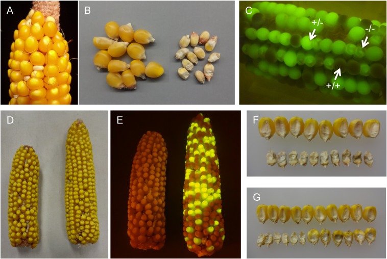
The dek38-Dsg mutant segregating in an ear (A, small kernels) and after shelling (B, Right). When viewed under a blue light (C), segregation between normal non-GFP (+/+), normal GFP (+/−), and dek GFP (−/−) is visible. The change in color of normal kernels not fluorescing GFP is because of exposure to blue light in a dark background. (D, Left) Ear from a dek38 footprint allele with with a frameshift mutation. (D, Right) Ear from the allelism test between the same footprint allele and the dek38-Dsg parent. (E) Same ears as in D, but under blue light. The frameshift allele on left lost Dsg (and fluorescence), but the kernels on the right fluoresce because of the Dsg element in the dek38-Dsg mutant. Also shown are pictures of defective kernels (embryo side up) from the footprint allele (F) and from the allelism test (G). Normal kernels are shown on top for comparison.

