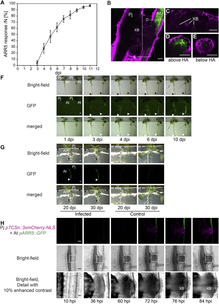
Fig. S4.
Phtheirospermum induces pARR5::GFP response in Arabidopsis. (A) The ratio of Arabidopsis pARR5::GFP plants showing GFP expression above haustoria (ARR5 response) was quantified between 3 and 11 dpi in four independent experiments with each containing between 34 and 46 infected plants (mean ± SD). (B) Z projection of 17 images separated by 2 µm of a two-photon microscope scanned Phtheirospermum (Pj) haustorium on Arabidopsis pARR5::GFP (At) at 11 dpi with indicated optical cross-section through the haustorium (C), the Arabidopsis root above (D) and below (E) the haustorium. Propidium iodide-stained cell walls are shown in magenta, GFP in green, XB (xylem bridge). Images of At pARR5::GFP plants infected with Pj were taken at indicated time points during cultivation in infection media (1–10 dpi) (F) and on growth media (1/2 MS agar, 20 dpi and 30 dpi) (G). Arrows indicate haustorium attachment sites. (H) Fluorescent and bright-field images as shown in Fig. 2D of Pj hairy roots expressing pTCSn::3xmCherry-NLS (magenta) and infecting host roots (At pARR5::GFP, green) with detailed images of the parasite–host interface as indicated by dotted lines in the bright-field panel. (Scale bars: B–E, 50 µm; F and G, 1 mm; H, 100 µm.)

