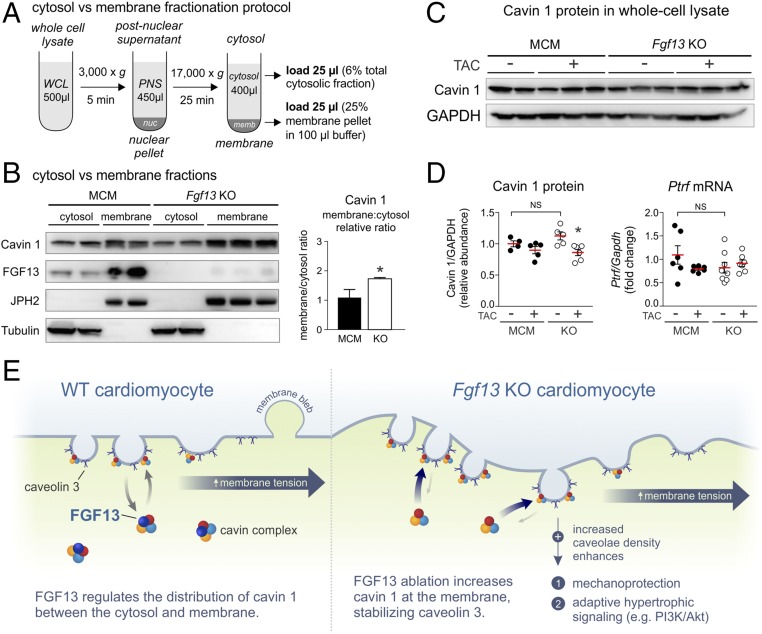
Fig. 7.
KO of Fgf13 in adult cardiomyocytes redistributes cavin 1 from the cytosol to membrane. (A) Schematic of subcellular fractionation workflow (PNS, postnuclear supernatant; WCL, whole-cell lysate). Equal concentrations of lysate were used as input. (B, Left) Western blots for cavin 1 and FGF13 from cytosolic and membrane fractions prepared from MCM and Fgf13 KO ventricular tissue; tubulin and junctophilin-2 (JPH2) serve as markers for cytosolic and membrane proteins, respectively. (Right) Quantification of membrane:cytosol relative ratio for cavin 1. *P < 0.05, unpaired t test. (C) Representative Western blots for cavin 1 in left ventricular whole-cell lysates from MCM and Fgf13 KO hearts 12 wk post-TAC or sham surgery. (D, Left) Densitometry of cavin 1 protein from blots in C. (Right) Gene expression of cavin 1 (Ptrf). *P < 0.05 vs. KO sham, two-way ANOVA with Sidak’s multiple-comparison test. (E) Model showing FGF13 regulation of cavin 1 distribution between cytosolic and membrane fractions. Fgf13 KO increases cavin 1 at the membrane, stabilizing caveolin 3 and thereby increasing caveolae density to enhance mechanoprotection and adaptive hypertrophic signaling.

