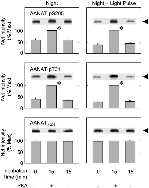
Fig. 2.
Light exposure reduces the degree of phosphorylation of T31 and S205. The percentage of phosphorylated AANAT pT31 and AANAT pS205 in ovine pineal glands at night (Left) and after exposure to 30 min of light at night (Right) was determined. AANAT was maximally phosphorylated by incubation with ATP and PKA. Each sample was analyzed in duplicate on a 15% gel containing the three treatment groups indicated; there was no decrease in phosphorylation during incubation in the absence of PKA and ATP. The data on the left were not normalized to the data on the right. Each digital image is an example of one sample representative of the experimental group. Each bar represents the mean ± SE of the results of analysis of three samples (two glands per sample). For additional details, see Materials and Methods and the Fig. 1 legend. *, Incubation with PKA and ATP increased the degree of phosphorylation (P < 0.01).

