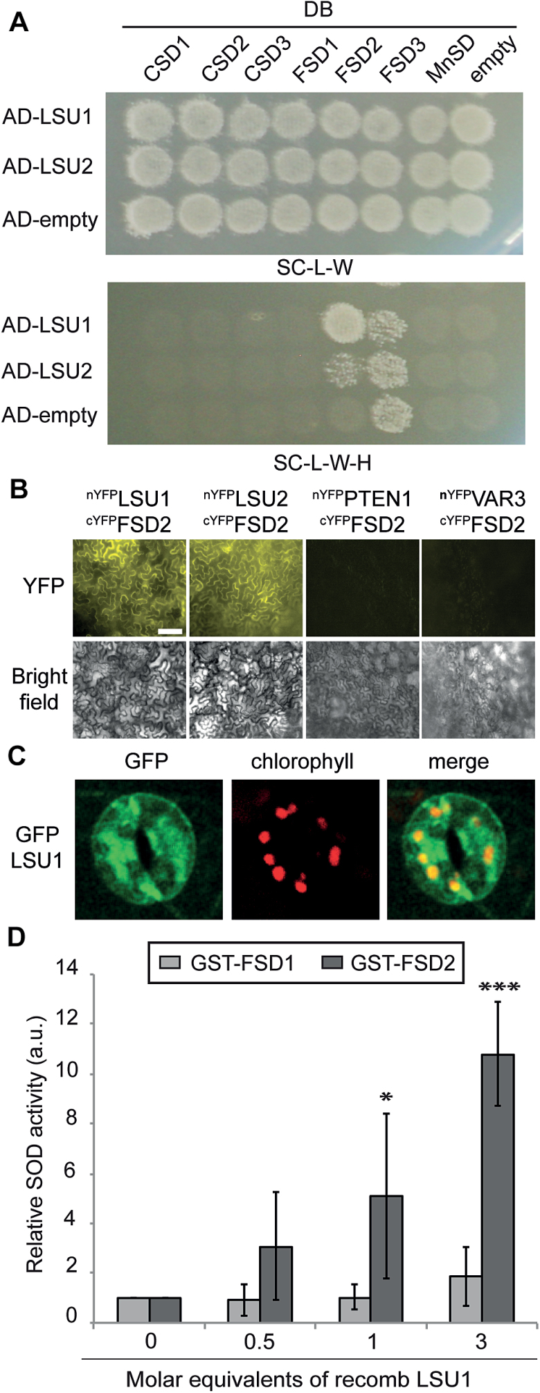
Fig. 4.
LSU1 and LSU2 physically and functionally interact with FSD2. (A) Specific interaction of AD–LSU1 and AD–LSU2 with DB–FSD2 by Y2H. The top panel shows diploid yeasts and successful mating; the bottom panel shows growth on selective media indicating interactions of AD–LSU1/2 with DB–FSD2 and autoactivation of DB–FSD3. (B) Bimolecular fluorescence complementation (BiFC). Nicotiana benthamiana epidermal leaves transiently co-expressing cYFP–FSD2 and nYFP–LSU1/2 restore YFP fluorescence, whereas co-infiltration of cYFP–FSD2 and nYFP fusions with the cytosolic Arabidopsis PHOSPHATASE AND TENSIN HOMOLOG DELETED ON CHROMOSOME TEN 1 (PTEN1, AT5G39400) and with the chloroplast-localized nYFP–VARIEGATED 3 (VAR3, AT5G17790) do not (scale bar=100 µm). (C) LSU1 partly co-localizes with guard cell chloroplasts. Representative confocal images of guard cells in first leaflets of –S-grown GFP–LSU1 seedlings. Shown are GFP signal (left), chlorophyll autofluorescence (middle), and merged channels (right) (scale bar=25 µm). (D) Relative in vitro SOD activity of recombinant GST–FSD1 and GST–FSD2, respectively, in the presence of increasing concentrations of MBP–LSU1. Error bars correspond to the SD of three independent experiments, and significant differences from controls were assessed using Student’s t-test (*P<0.05; ***P<0.001).

