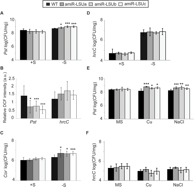
Fig. 6.
LSU proteins mediate defence responses during abiotic stress. WT and amiR-LSUa–c seedlings were flooded with bacterial suspension at 5 × 10⁶ CFU ml–1 of the P. syringae strains: Pst DC3000 (Pst), COR–, and hrcC. In (A) and (C–F), colony-forming units were determined 3 days post-infection (dpi) and normalized to fresh weight. Seedlings were grown in normal (+S) and –S conditions, or 1/2 MS conditions, or submitted to high Cu or NaCl, as indicated. (B) DCF fluorescence in guard cells of –S-grown lines at 1 dpi with the indicated bacterial strain normalized to mock treatment. Error bars correspond to the SD of at least three biologically independent experiments, and statistical differences from the WT were assessed using Student’s t-test and are indicated with asterisks (*P<0.05; **P<0.01; ***P<0.001).

