. 2017 Apr 7;6:e24665. doi: 10.7554/eLife.24665

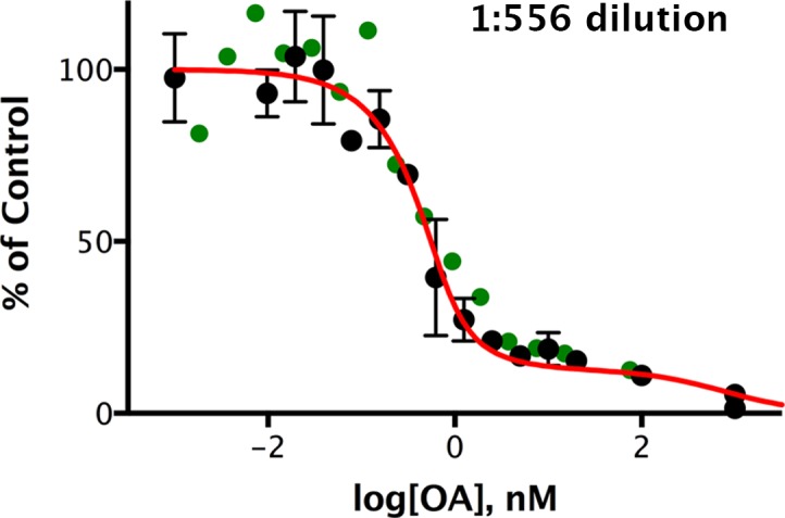
Figure 7. Okadaic acid inhibition of the dephosphorylation of pCPI-17 by dilute mouse uterus extracts.

Mouse uterus extracts at a dilution of 1:556 were prepared as described in Materials and methods and incubated at 30°C for 45 s with 687 nM 32P-labeled pCPI-17 plus the indicated concentrations of okadaic acid (OA). The mean phosphate released (with standard deviations; n = 3) is shown as a percentage of release in the absence of OA. Since the pCPI-17 concentration is much greater than the MLCP concentration at these dilutions, essentially all dephosphorylation is due to PP2A and other non-MLCP phosphatases (collectively called PPU).
DOI: http://dx.doi.org/10.7554/eLife.24665.012

Figure 7.

Figure 7—figure supplement 1. Okadaic acid inhibition of the dephosphorylation of pCPI-17 by dilute mouse uterus extracts.
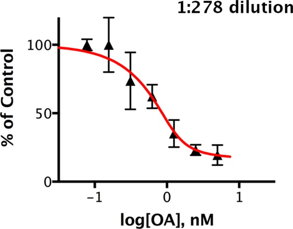
Figure 7—figure supplement 1.

This experiment is identical to that shown in Figure 7 except that the mouse uterus extracts were diluted 1:278 and incubated at 30°C for 30 s with 32P-labeled pCPI-17 plus the indicated concentrations of okadaic acid (OA). The mean phosphate released (with standard deviations; n = 3) is shown as a percentage of release in the absence of OA.
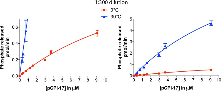
Figure 7—figure supplement 2. Kinetic analysis of the dephosphorylation of pCPI-17 by PPU.

Figure 7—figure supplement 2.

Highly diluted uterus extract was incubated with the indicated concentrations of 32P-labeled pCPI-17 for either 30 s (30°C) or 3 min (0°C) in 4 μl reactions. Values and standard deviations are for three experiments at each temperature.