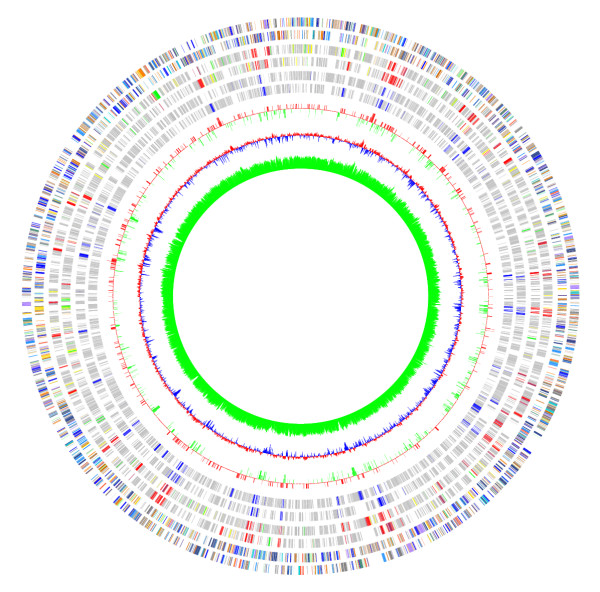
Figure 3.

Comparing data from different sources using GenomeViz.The figure shows a comparison of the distribution of horizontally transferred genes in Escherichia coli K12 compiled from three different sources. From outside to inside: Escherichia coli K12 COG categories (two circles), genes identified by SIGI (two circles), genes listed in the Horizontal Gene Transfer Database [18] (two circles), standard deviations of genes identified by IslandPath (single circle, red +ve, green -ve), mean centered GC content of the genome (red: above mean, blue: below mean), GC content of the genome again as a single-sided line plot (green).
