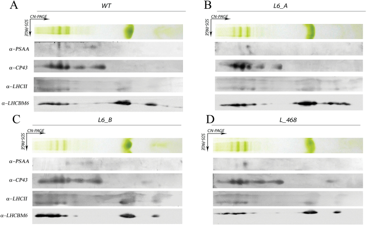
Fig. 5.
Analysis of the thylakoid membrane pigment–protein complexes by 2D electrophoresis and immunoblotting. Thylakoid membranes of knock-down strains grown in control light were solubilized with 1% dodecyl-maltoside (α-DM) and separated by CN–PAGE followed by a second dimension separation by SDS–PAGE. Immunoblot detection of LHCBM4/6/8, LHCII, PSI (antibody α-PSAA), and PSII (antibody α-CP43) is also reported. (This figure is available in colour at JXB online.)

