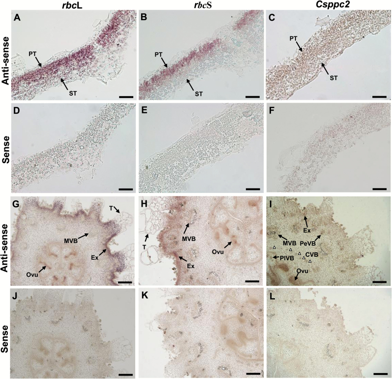
Fig. 4.
In situ hybridization of rbcL, rbcS, and ppc transcripts in cucumber fruits. (A–F) Leaf cross-sections (0–1 DAU) hybridized with the rbcL, rbcS, and Csppc2 antisense (A–C) and sense (D–F) probes, respectively. (G–L) Young ovary/fruit cross-sections (–2 –0 DAA) hybridized with the rbcL, rbcS, and Csppc2 antisense (G–I) and sense (J–L) probes, respectively. White triangles in (I) indicate the CVB. CVB, carpel vascular bundle; Ovu, ovule; PeVB, peripheral vascular bundle; PlVB, placenta vascular bundle; T, trichome; for other abbreviations, see Figs 1 and 2. Scale bars=50 µm in (A–F) and 200 µm in (G–L).

