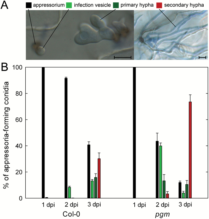
Fig. 1.
Colletotrichum higginsianum hyphae proliferation in pgm. Leaves of 5-week-old plants were infected with C. higginsianum, stained with trypan blue at 1, 2, and 3 d post infection (dpi) and examined by differential interference contrast microscopy. (A) Micrographs illustrating the scored fungal infection structures. Bars represent 10 µm. (B) Early fungal in planta development as given by the relative distribution of infection structures. Values are means of three biological replicates with four individual leaves each. Per replicate, the developmental status of in planta hyphae formed from 400–500 conidia was scored. The error bars represent the SE. Starting from appressoria, the most advanced infection structure was classified. The developmental order is as follows: appressoria, black bars; infection vesicles, light green bars; primary hyphae, dark green bars; secondary hyphae, red bars.

