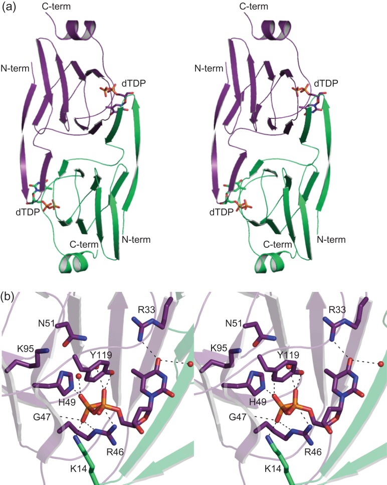
Fig. 4.
The structure of WlaRA. Shown in stereo in (A) is a ribbon representation of the WlaRA dimer. Subunits 1 and 2 are highlighted in violet and green, respectively. The bound dTDP molecules are displayed in stick representations. A close-up stereo view of the active site in subunit 1 is presented in (B). Ordered water molecules are represented by red spheres. Possible hydrogen bonding interactions, within 3.2 Å, are indicated by the dashed lines. Due to domain swapping, Lys 14 is contributed by subunit 2. This figure is available in black and white in print and in color at Glycobiology online.

