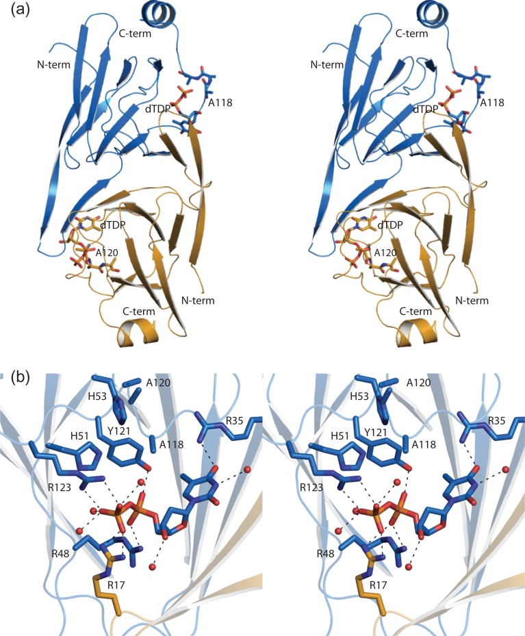
Fig. 5.
The structure of WlaRB. A stereo ribbon representation of WlaRB is presented in (A) with subunits 1 and 2 color coded in blue and yellow, respectively. The positions of the site-directed mutations made in order to produce X-ray diffraction quality crystals are shown (E118A, K119A and E120A) in stick representations. The dTDP ligands are also displayed in stick representations. A close-up stereo view of the WlaRB active site is presented in (B). Arg 17 is contributed by subunit 2 in the dimer. Possible hydrogen bonding interactions are indicated by the dashed lines. This figure is available in black and white in print and in color at Glycobiology online.

