
Fig. 5. Modeling MTs in the contracting sarcomere.
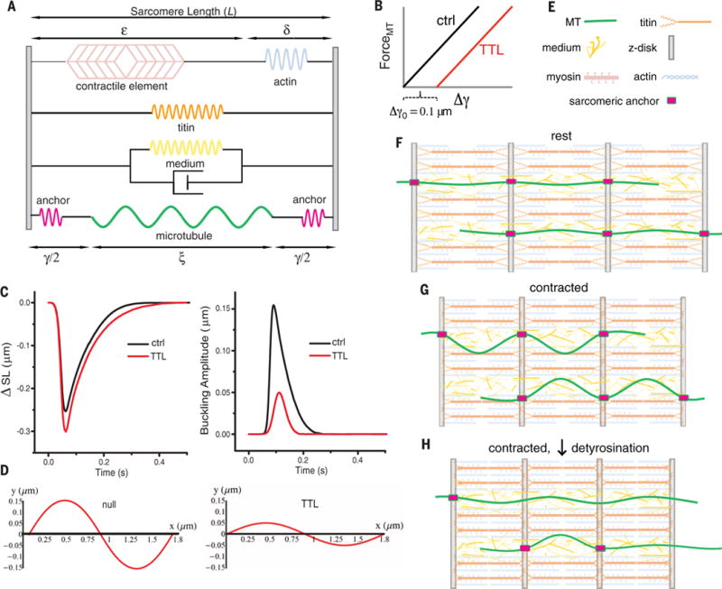
(A) Mechanical schematic of the modeled sarcomere. A force-generating contractile arm (top) is coupled in parallel at the Z-disc to a spring element representing titin (orange), a viscoelastic medium (yellow spring and dashpot), and a MT (green) with anchors (fuschia pink) to the Z-disc (gray). The anchor to the Z-disc is only engaged at regions of MT detyrosination. (B) TTL overexpression is modeled by allowing the anchors to slide for 50 nm at each end before engaging and transmitting force to the MT at a detyrosinated subunit. (C) The change in sarcomere length at peak contraction and buckling amplitude. (C) and (D) recapitulate experimental observations for TTL-overexpressing myocytes after this change. (E and F) The cardiac sarcomere, shown with MTs with putative stiff anchors to the sarcomere, here, at the Z-disc. Contraction reduces the distance between anchor points, which requires the MTs either to buckle (G) if the anchors are engaged or to slide (H), if the anchors are not engaged and force incident on the MT remains low. Mathematical model parameters are available in table S5.