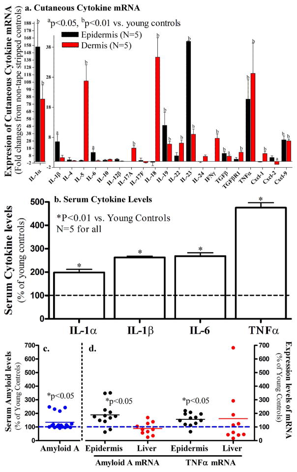
Figure 2. Cutaneous and Serum Cytokines Increase in Aged Mice.
Both flanks of 12-month old C57BL/6J mice were used in this study. The dermis and epidermis were separated by heat (Feingold et al., 1991). Fig 2a: Expression levels of cytokine mRNA in the skin; Fig 2b: Serum cytokine levels; Fig 2c: Serum amyloid A levels in 7-week old vs. 12-month old mice. Fig 2d: mRNA levels for amyloid A and TNFα in the liver (red dots) and the epidermis (black dots) of 12-month old mice. Data were normalized to normal young controls, setting normal young controls as 100% (dotted lines). A Mann Whitney two-tailed test was used to determine the significances between aged and young mice. P values were vs. the non-tape stripped normal controls.

