
Figure 6. Impaired RPE-cone interaction and photoreceptor alignment in canine bestrophinopathy.
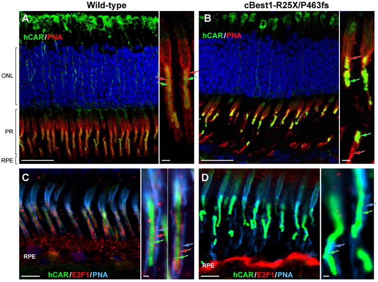
(A, B) Confocal photomicrographs of canine wild-type (A) and cBest1-R25X/P463fs-affected (B) retinae immunolabeled for human cone arrestin (hCAR, green) and PNA lectin (red). PNA-positive cone extracellular matrix sheaths tightly embrace the E2F1-labelled cone-specific RPE projections, extending along the outer- and toward the inner segment of cone photoreceptor cells in the control retina (A, close-up); in the cBest mutant, this IPM domain appears ruptured, exposing OS to the extracellular space (B, close-up). Together with the impairment in structural integrity of cone-specific IPM escheatment, cone photoreceptor OS lose their proper orientation (B), necessary for the normal interactions with RPE cells. (C, D) Multi-label IHC images of canine wild-type (C) and cBest mutant (D) retina illustrating bilayered extracellular compartmentalization of the cone-specific escheatment responsible for a normal apposition of RPE-cone OS (C, inset). This well-defined extracellular structure was lost in the affected retinae (D), revealing a dearth of the intrinsic RPE-associated apical microvillus layer, accompanied by rather fragmented external insoluble cone-specific matrix domain (D, close-up). Cone OS stripped of their protective sheaths appeared undermined with no direct contact with the underlying RPE (B, D). hCAR: human cone arrestin (shown in green A-D, and delineated by green arrows on the magnified images); PNA: peanut agglutinin lectin (shown in red A-B, and defined by red arrows on the corresponding close-ups; PNA in C-D is represented in cyan, and by cyan-colored arrows on the magnified photomicrographs); E2F1: E2F transcription factor 1 (shown in red C-D, and indicated by red arrows in C inset). ONL: outer nuclear layer; PRs: photoreceptors; RPE: retinal pigment epithelium. Scale bars: 20µm (A, B), 10µm (C, D), and 1µm (applies to all magnified images). Photomicrographs C & D: modified from Guziewicz et al. Adv. Exp. Med. Biol. (in press).