
Figure 8. EZRIN immunolabeling in WT, BVMD, and ARB hiPSC-RPE.
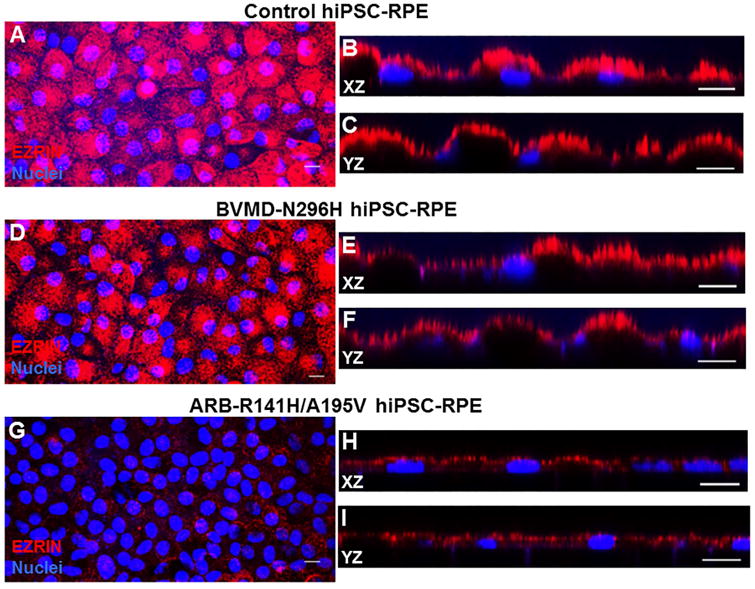
(A-C) Immunohistochemical analysis of WT hiPSC-RPE showed strong expression of EZRIN with vertically oriented, apical localization. (D-F) Similar expression and localization of EZRIN was observed in BVMD hiPSC-RPE. (G-I) In the ARB hiPSC-RPE, harboring compound heterozygous mutation in BEST1, MV are smaller in height compared to the WT hiPSC-RPE, and express EZRIN in a punctate fashion within a thin apical band. XZ and YZ labeled images are representative images for X-Z and Y-Z optical sections. Scale bar: 10µm applies to all panels.