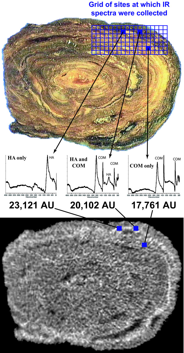
Figure 2.
Calibrating micro CT attenuation to pure mineral. On stone slice shown (top) FT-IR spectra were collected on each of 126 regions indicated by the grid area. Examples of spectra are shown for three regions, which represent the three classes of spectra collected from cut surface of this stone. Spectra indicated that the mineral was either purely calcium oxalate monohydrate (COM), purely apatite (HA), or a mixture of these two minerals. Corresponding regions-of-interest on micro CT image slice (bottom) taken just beneath the cut surface are shown with blue squares, and micro CT attenuation values are given below spectra. Using this method, regions of pure mineral were identified on stone slices and corresponding regions-of-interest measured in micro CT images to determine CT attenuation of different minerals.

