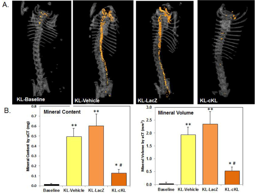
Figure 1. AAV-cKL effects on aortic calcification.
(A) Representative uCT images of aortic calcification (orange colorization) from αKL-null (KL) mice at baseline (four weeks of age), AAV-cKL treated, as well as vehicle and AAV-LacZ controls (treated from four weeks of age for four additional weeks). cKL administration was associated with a visually marked reduction in aortic mineralization versus KL-vehicle and KL-LacZ treated mice. (B) Mineral content and mineral volume of whole aortae were quantified and determined to be significantly elevated in controls (**p<0.01 and *p<0.05 vs baseline), whereas in cKL-treated mice mineral content and volume were significantly reduced (#p<0.005). [From: Hum JM, O’Bryan LM, Tatiparthi AK, Cass TA, Clinkenbeard EL, Cramer MS, Bhaskaran M, Johnson RL, Wilson JM, Smith RC, White KE. Chronic hyperphosphatemia and vascular calcification are reduced by stable delivery of soluble klotho. JASN (2016).]

