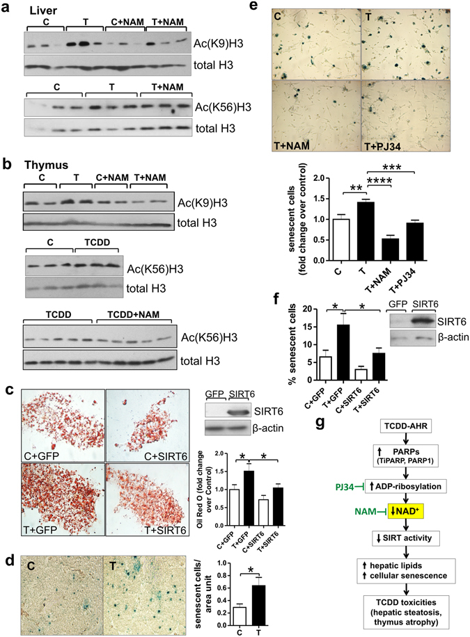
Figure 4.

NAD+ loss by TCDD decreases Sirt6 activity. Role for Sirt6 in TCDD toxic effects. (a) Western blots on homogenates of liver and thymus from CE treated with TCDD or vehicle (n = 3 independent experiments) for 24 hr. CE were cotreated with class I/II deacetylase inhibitors as described in Methods, to allow H3K9 and H3K56 acetylation to serve as indices of Sirt6 deacetylase activity. (b) Western blots on homogenates of liver and thymus from CE treated with TCDD (24 hr) with or without NAM for the last 4 hr. (c) Representative images of Oil red O stained CEH transfected with an empty plasmid (GFP) or a plasmid containing chick Sirt6 and treated with TCDD (T) or solvent (C). Upper right panel: Western blot on transfected CEH homogenates. (d) Representative images for SA-Xgal stained sections of thymus glands from CE treated with TCDD or vehicle for 48 hr. Bar graph shows means ± SE for numbers of senescent cells (blue stained) per unit area of thymus. (e) SA-Xgal stained thymic epithelial cells from CE treated with TCDD for 48 hr with or without NAM or PJ34, each injected right after TCDD treatment and 24 hr later. Bar graph shows the mean incidence of senescent cells ± SE per treatment group. (f) Means ± SE for incidence of senescent (SA-Xgal stained) thymic epithelial cells transfected with pcDNA-GFP or pcDNA-Sirt6 and treated for 48 hr with TCDD or vehicle. Right panel: Western blot on homogenates of thymic epithelial cells transfected with GFP or Sirt6 plasmids. (g) Scheme showing the proposed mechanism by which increased PARP activity produces NAD+ loss leading to diverse toxic effects of dioxin in vivo.
