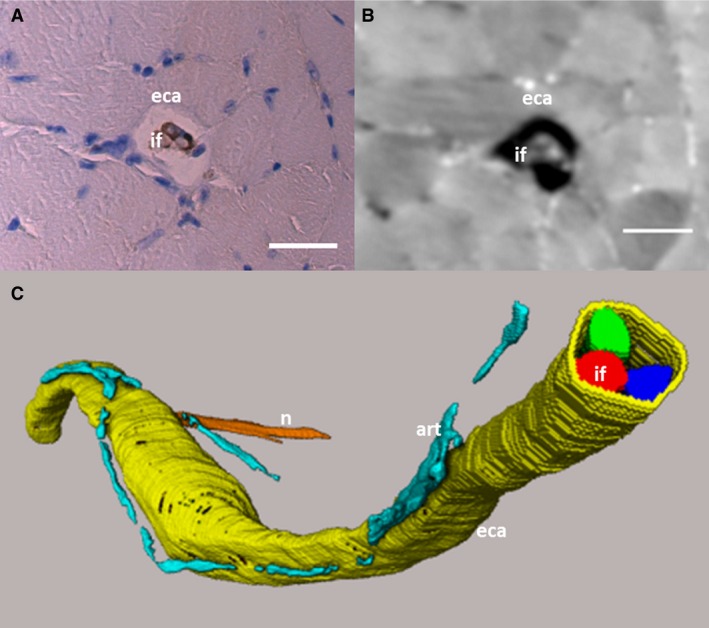
Figure 2.

Histological and SR CT slice of muscle spindle and 3D volume rendering. (A) Histological slice corresponding to (B) the SRCT image. The external capsule (eca) and intrafusal fibres (if) can be distinguished in the SR CT image. Scale bars: (A,B) 30 μm. (C) The different intrafusal fibres (red, blue, green) and the external capsule (yellow) have been segmented from the SR CT data, along with the lumen of small arterioles (art, turqoise) running along the spindle and one of the supplying nerves (n, orange). The segmented data is visualised through volume rendering. The S‐shape of the muscle spindles is due to the muscle not being stretched during fixation, which led to a distortion of the muscle.
