
Figure 6.
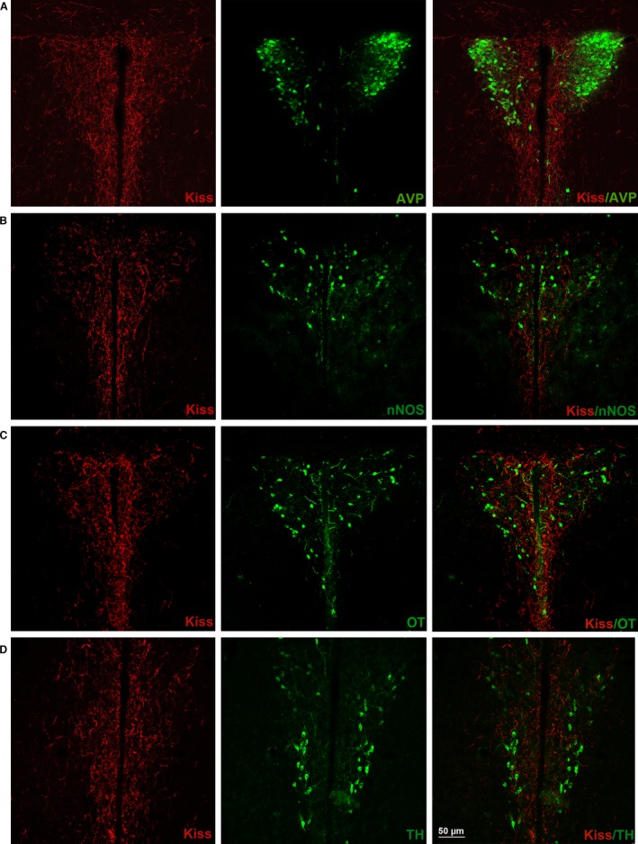
Kisspeptin (Kiss) and paraventricular nucleus (PVN) cellular populations [arginine vasopressin (AVP), neural nitric oxide synthase (nNOS), oxytocin (OT), tyrosine hydroxylase (TH)]. Coronal section of the adult CD1 female mice PVN in estrus phase. It is possible to observe the relations with Kiss (red) and different PVN neuronal populations: (A) AVP (green); (B) nNOS (green); (C) OT (green); (D) TH (green). Note that the total AVP and the majority of nNOS neuronal cell bodies were distributed in lateral PVN, where the concentration of Kiss fibers was lower; in the medial PVN instead, a conspicuous number of OT and TH cell bodies were present.