Fig. 1.

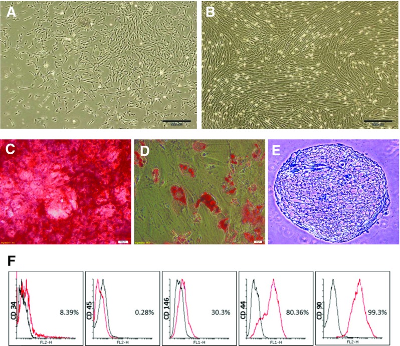
Dental pulp stem cells (DPSCs). a In primary cultures, the cells formed large colonies that consisted of fibroblastic cells (bar = 200 μm). b The colonies increased in size, becoming confluent (bar = 200 μm); the shiny dots were other cells that are beside dental pulp cells in pulp chamber and cultured with them. c Osteogenic differentiation of DPSCs stained by Alizarin Red (bar = 100 μm). d Adipose differentiation of DPSCs stained by Oil Red O (bar = 20 μm). e Cartilage differentiation of DPSCs stained by toluidine blue (bar = 100 μm). f The cells had a surface antigenic profile similar to those of mesenchymal stem cells. While endothelial and hematopoietic markers were present in a very low percentage of the cells, the mesenchymal markers were expressed by the majority of the cell population. FITC Z fluorescein isothiocyanate, PE Z phycoerythrin
