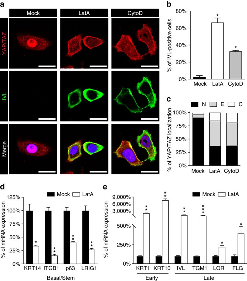
Figure 3. F-actin preserves epidermal stemness by sustaining YAP/TAZ and opposing differentiation.
(a) IF microscopic images of nHEK cells treated for 24 h with vehicle (Mock), 0.8 μM latrunculin A (LatA) or 2.5 μM cytochalasin D (CytoD). Images show the endogenous YAP/TAZ proteins (red) and the Involucrin differentiation marker (IVL, green). DAPI (blue) is a nuclear counterstain. Scale bar: 20 μm. (b) Quantitation of Involucrin expression after treatment for 24 h with actin inhibiting drugs as in a. Bars represent mean+s.d. (n=3 independent experiments. *P<0.0001 compared to Mock-treated cells; Student's t-test). (c) Proportion of YAP/TAZ subcellular distribution in nHEK cells treated with the indicated drugs and analysed as previously described. Bars represent mean of three independent experiments. (d,e) nHEK cells treated for 24 h with vehicle (Mock) or 0.8 μM latrunculin A (LatA) were analysed by qRT–PCR for the expression of either basal and stem marker genes (KRT14, ITGB1, p63, LRIG1) (d) or early (KRT1, KRT10) and late (IVL, TGM1, LOR, FLG) differentiation genes (e). Notably, LatA potently promoted differentiation as shown by the upregulation of the indicated markers. At the same time, disrupting F-actin integrity is sufficient to reduce the expression of basal (KRT14) and SC (ITGB1, p63 and LRIG1) markers. Bars represent mean+s.d. *P<0.05, **P<0.01, ***P<0.001 compared to Mock; Student's t-test). See Methods section for reproducibility of experiments. See also Supplementary Fig. 2g for the effect of F-actin-inhibiting drugs on YAP/TAZ transcriptional activity in keratinocytes.

