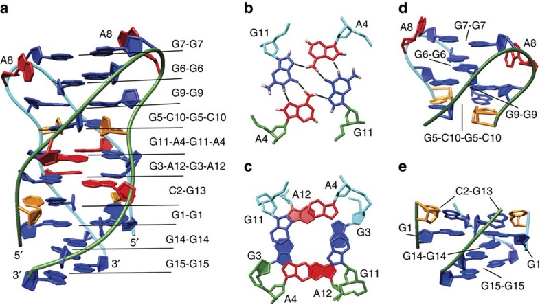
Figure 3. Structure of VK34 dimer.
(a) Representation of the lowest-energy overall structure. (b) Top view of the G11-A4-G11-A4 quartet with marked hydrogen bonds. (c) Top view of stacked G3-A12-G3-A12 and G11-A4-G11-A4 quartets with a helical twist of 25° depicted with filled and unfilled residues, respectively. (d) Side view of the G5-C10-G5-C10 quartet and G6-G6, G7-G7 and G9-G9 base pairs. (e) Side view of two buckled C2-G13 base pairs and G1-G1, G14-G14 and G15-G15 base pairs. The guanine residues are coloured blue, adenine red and cytosine orange. The two strands are coloured green and cyan.

